Bottom
Previous
Contents
The Braggs and Astbury:
Leeds and the beginning of Molecular Biology
Anthony North, Astbury Professor of Biophysics (Emeritus)
Astbury Centre for Structural Molecular Biology,
The University of Leeds
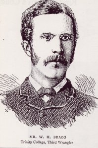
William Henry Bragg was born in Wigton, Cumbria in 1862 and graduated in
Mathematics from Cambridge in 1885. He came 3rd in the final examinations (a
position known as 3rd Wrangler). In his final year he had done some work in
the Physics department and become known to J J Thomson (later a Nobel
Laureate for his discovery of the electron); shortly after graduating he was
encouraged by Thomson to apply for the position of Professor of Mathematics
& Physics at the University of Adelaide. He was duly appointed to the
post and set sail for Australia - the lengthy sea voyage gave him the
opportunity to read up some more physics!
W H Bragg in Adelaide
On his appointment in 1886, he found that, with just one assistant, he was
entirely responsible for all the mathematics and physics teaching in the
University, adding up to 18 hours per week plus 6 hours evening FE classes.
In order for his students to do practical work, he apprenticed himself to a
firm of instrument makers to make apparatus for his classes - and his
interest in instrumentation was to prove important in later years.
But it was not all hard work - he was an excellent tennis player and joined
in the social life of Adelaide. He met Charles Todd, who held the posts of
South Australian Government Astronomer, Postmaster-General and
Superintendent of Telegraphs. Todd had been responsible for constructing the
telegraph line from Adelaide to Darwin and had named the staging post 'Alice
Springs' after his wife. WHB got to know Todd's family and in 1889 he
married Gwendoline Todd, with whom he had 3 children, William Lawrence,
Robert and Gwendolen.
|
|
Initially, he had little opportunity for research, (and, indeed, said in
later years that it had never occurred to him to do any) but he was very
interested in the developing ideas about electromagnetism, wireless and,
particularly, radioactivity; and he constructed apparatus for demonstrating
these phenomena to his students.
It was not until 1903 that WHB carried out his first serious independent
research; he acquired some radium and carried out experiments on
radioactivity, alpha & beta particles and gamma rays.
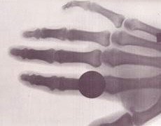 X-ray of Bragg's hand
|
Röntgen had discovered X-rays in 1895, but their nature was the subject
of controversy. WHB took X-ray pictures of his own hand and of his son's
elbow, hurt in a cycling accident. At this time, there was controversy about
the nature of X-rays - were they particles or waves?
WHB became a proponent of the idea that X-rays were particles
and he had detailed correspondence with Rutherford and others. He became
known internationally and was elected FRS in 1907. He had, however, become
concerned that Adelaide was remote from world-leading laboratories and
considered a move; the possibility of succeeding Rutherford at McGill
University in Canada was frustrated by a serious fire there, but in 1908, he
was invited to the Cavendish Chair of Physics in Leeds succeeding Professor
William Stroud.
|
W H Bragg in Leeds
WHB and his wife were initially unhappy in Leeds; the city was dirty with
poor housing and his research seemed to have stagnated. He continued to be
engaged in a controversy with Barkla as to whether X-rays were waves or
corpuscles. (In retrospect, Bragg had been studying high-energy rays and
Barkla softer, low-energy rays, so they had been observing different
properties.)
However, all was to change in 1912, when von Laue, Friedrich & Knipping
shone X-rays on to crystals and obtained a pattern of spots on photographic
film. But, were the spots on the film due to corpuscles passing through
channels between the atoms in the crystal or to waves diffracted by the
atoms?
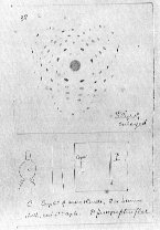
WHB wrote to Arthur Schuster enclosing this diagram.
"I enclose a drawing of the curious X-ray effect obtained by Dr Laue in
Munich. It is claimed, I understand, that it is a diffraction effect due to
the regular arrangement of the molecules in space"
|
|
Cambridge
WHB's elder son (WLB) had studied in his father's department in Adelaide,
but he had moved to England with the family in 1909 and, in 1912, he
graduated in physics in Cambridge.
The Cavendish Laboratory, Cambridge, 1913
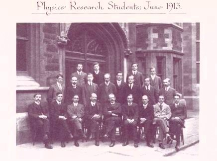
WD Rudge RW James WA Jenkins JK Robertson
WL Bragg VJ Pavlov S Kalandyk FW Aston HA McTaggart H Smith F Kerschbaum AN Shaw
RD Kleeman AL Hughes R Whiddington CTR Wilson JJ Thomson F Horton RT Beatty AE Oxley G Stead
R Whiddington became Professor of Physics in Leeds, RW James
became a
leading crystallographer in South Africa, and apart from WL Bragg were three
other Nobel Prize winners: FW Aston became known for his work on isotopes
and received the Nobel Prize for Chemistry in 1922, JJ Thomson received the
Nobel Prize for the discovery of the electron, CTR Wilson received the 1927
Nobel Prize in Physics for his invention of the cloud chamber.
While in Cambridge, WLB was of course intrigued by the results of Laue,
Friedrich and Knipping, for which they had produced a complicated
explanation and, in late 1912, he had his 'brain wave': the pattern of spots
in the X-ray pattern from a crystal could be explained by 'reflection' of
waves from crystal planes, analogous to light rays from a mirror.
He published his idea in a paper entitled "The diffraction of short
electromagnetic waves by a crystal" in Proceedings of the Cambridge
Philosophical Society in late 1912. The title was carefully chosen - he
did not want directly to contradict his father's view that X-rays were
actually particles. The facilities for doing experiments with X-rays in
Cambridge were rather poor, whereas WHB had constructed an X-ray
spectrometer in Leeds, which gave much more reliable measurements than the
Cambridge cameras and films. During the vacations, WLB worked in his
father's labs and during 1912-13, the 2 men worked together, observing
X-rays reflected from cleaved mica and then deriving the arrangement of
atoms in simple inorganic crystals, including NaCl and KCl.
An aside - Sodium Chloride
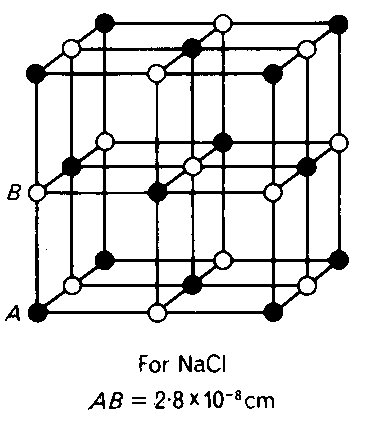 |
For a long time, chemists refused to accept the fact that NaCl contains no
NaCl molecules, the crystals containing just an alternating array of
Na+ and Cl- ions. WLB used to recall chemists trying
to persuade him that the Na+ and Cl- ions were not
exactly equidistant from each other, but that they might be arranged in
pairs just a little closer to each other!
Even as late as 1927, Professor H E Armstrong wrote in a letter to Nature:
"Professor W L Bragg asserts that in Sodium Chloride there appear to be
no molecules represented by NaCl. The equality in number of sodium and
chlorine atoms is arrived at by a chess-board pattern of these atoms; it is
a result of geometry and not a pairing-off of the atoms.
"This statement is more than 'repugnant to common sense'. It is absurd to
the nth degree, not chemical cricket. Chemistry is neither chess nor
geometry, whatever X-ray physics might be. Such unjustified aspersion of the
molecular character of our most necessary condiment must not be allowed any
longer to pass unchallenged. It were time that chemists took charge of
chemistry once more and protected neophytes against the worship of false
gods; at least taught them to ask for something more than chess-board
evidence."
|
1914 and onwards:
UCL, Manchester and the Royal Institution
During the first world war, WHB became much involved with scientific advice
to the government and decided to move from Leeds to University College,
London, to be nearer the centre of national activity. WLB was on active
service in France developing acoustic range-finding methods for directing
gunfire and was invested with the Military Cross. Sadly, his younger brother
Robert was killed in action.
In 1915, WHB and WLB were awarded the Nobel Prize. This is still the only
example of the joint award of a Nobel prize to father and son and WLB
remains the youngest recipient of a Nobel prize at the age of 25.
After the war, WHB and WLB continued their X-ray diffraction studies, but
agreed to work on different aspects.
After a few months back in Cambridge, in 1919 WLB succeeded Ernest
Rutherford in the Langworthy Chair of Physics in Manchester, and for many
years he concentrated mainly on metals and minerals, especially the
silicates, and on the development of diffraction methods.
Meanwhile, WHB had built up a research team at University College London,
working largely on organic substances. While there, he wrote: "My son and
I have been comparing notes and we find that we can only get a few hours
each week for research" - an experience shared by most present-day
academics!
In 1923, WHB succeeded Sir James Dewar as Resident Professor and Director of
the Davy-Faraday Laboratory at the Royal Institution, where he would be free
from routine teaching. He took with him members of his UCL team, including
William Astbury and Kathleen Yardley (later Lonsdale). Other research
workers at the R.I. included J M Robertson (for many years a leading
chemical crystallographer in Glasgow), E G Cox (later Professor of
Structural Chemistry in Leeds and subsequently Director of the Agricultural
Research Council) and J D Bernal, who became an important innovator of
studies of biological structures in Cambridge and London.
At the R.I., WHB re-established the series of lectures to lay audiences that
had been started by Davy and Faraday and he asked Astbury to take an X-ray
diffraction picture of a human hair for a lecture on "the imperfect
crystallisation of common things". Astbury had been doing good
crystallographic work, notably preparing with Kathleen Yardley the first
edition of what was to become the crystallographers' 'bible', the
International Tables of crystal symmetry relationships. Taking the fibre
diagram of hair stimulated Astbury's interest in the structures of natural
polymers.
The Vice-Chancellor of the University of Leeds, James Baillie, had decided
in 1928 that the Department of Textile Industries needed to be enlivened by
the appointment of a Lecturer in Textile Physics and WHB was among the
people consulted about the appointment; he wrote to Professor Aldred Barker:
"I have a man here who might possibly make you the research scientist you
want - W T Astbury. He is a really brilliant man, has done some first class
work which is quoted everywhere .... He has considerable mathematical
ability and a very good knowledge of physics and chemistry .... He is very
energetic and persevering, has imagination, and, in fact, he has the
research spirit. Although not trained in the workshop he is sufficiently
skilful with his hands.
He is a most loyal colleague to me and I do not
want to lose him at all but it is good for these people to make a move from
time to time. He can lecture, though I do not call him a very good lecturer,
he can write a great deal better than he can speak."
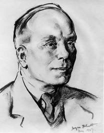
Despite the latter reservation, Astbury was appointed to Leeds as Lecturer
in 1928, where he was to spend the rest of his life, becoming Professor of
Biomolecular Structure in 1945.
Astbury in Leeds
 |
In those days, all textiles were made from naturally-occurring fibres such
as wool, silk and hair. Astbury set out to show that these were more than
the "biochemically lifeless and uninteresting material" they were often
claimed to be.
|
The Nature of Fibrous Materials
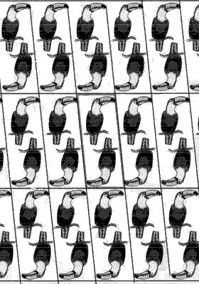
Normal crystals are made up from a regular pattern of 'unit cells', each of
an identical shape and content, which fill space in 3 dimensions. Each unit
cell contains one or more molecules, packed together in a symmetrical way.
In this example, each cell contains two 'molecules'.
We can only show one layer of 'molecules' in this picture - the complete
crystal would be formed by stacking layers one above the other.
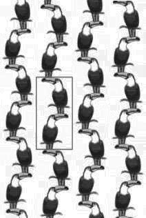
Fibrous molecules such as the biological macromolecules cellulose, wool,
silk and DNA too are very long and, in the fibre, are parallel to each
other, but they are not always lined up longitudinally with their neighbours
on either side - see below. We can, however, still have 'unit cells', with
the molecules passing through from one cell to the next along the chain,
each unit cell containing a number of the monomer units from which the chain
is composed. As with normal crystals, we still have regular lateral spacings
between unit cells and regular spacings along the chain, but we have lost
some of the regularity of a normal crystal when the chains are not in
register sideways. |
 |
Consequently, when an X-ray beam is shone at right angles to a fibre, the
'fibre diagram' obtained tends to show not the regular array of well-defined
spots as from a crystal, but a pattern of more diffuse spots and arcs.
Cellulose, the major structural polymer of plants, is a very simple
unbranched polymer of identical glucose units and the X-ray pattern from
cellulose - below - is therefore quite sharp.
|
 |
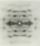 |
Successive glucose units, each 5.15 Å long, face in alternate
directions, like the toucans above, giving an exact repeat of 10.3 Å
along the fibre axis.
|
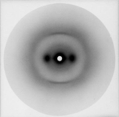
In contrast, the X-ray pattern from hair and wool, the 'alpha' pattern
(above left) is very diffuse, as they are proteins made up from a rather
irregular sequence of the 20 different types of amino acid. Nevertheless,
the positions of the spots and arcs give information about the geometry of
the chains and their spatial arrangement.
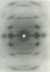
The pattern from silk, the 'beta' pattern (above right), is intermediate in
form because, although it too is a protein, there is greater regularity in
its amino-acid sequence.
Astbury found that, while wool and hair (both forms of the protein keratin)
normally gave the alpha type of pattern, they could be stretched in suitable
solvents to give a beta type pattern like that from silk. He made the
extremely important deduction that, in the alpha state, the molecules were
highly folded, but in the beta state they were stretched out.
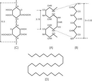
Measurements of the alpha pattern gave a longitudinal periodicity of
5.15Å - remarkably similar to half the periodicity (10.3 Å) of a
cellulose fibre. He found a periodicity of 9.96Å for the beta state.
Intrigued by the relationship of the alpha spacing to that of cellulose, he
proposed a model (A, below) for unstretched wool in which the protein chain
was bent back on itself in a shape rather like the glucose rings of
cellulose (C).
For the beta form, he put forward a model for the stretched-out protein chain.
D represents the 'cross-beta' form of protein chains, described later.
|
|
The Structure of Protein Chains
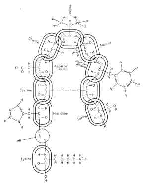
Protein molecules are polymers of 20 different kinds of monomer - the amino
acids. As the chain is synthesised on the ribosome, each additional amino
acid is added to the carboxyl end of the growing chain with the elimination
of a water molecule (bottom right).
The amino-acid side chains have widely differing sizes and properties:
- positively charged, e.g. lysine
- negatively-charged, e.g. aspartic acid
- neutral polar, e.g. serine
- non-polar aliphatic, e.g. alanine, valine
- non-polar aromatic, e.g. phenylalanine
- cystine, which cross-links two different parts of the chain,
stabilises (but complicates) the overall chain fold.
|
|
Astbury's classification of fibrous proteins
Probably his major contribution to the field was Astbury's wide studies of
the mechanical properties, physical properties (such as density) and X-ray
diagrams of as many naturally occurring fibrous polymers as he could find,
which led to the following generally accepted classification:
- collagen
- (the material of tendons, and supporting matrix of bone and skin)
- the k-m-e-f group:
- keratin (wool, hair and silk)
- myosin (one of the major muscle components)
- epidermin (a skin component)
- fibrinogen(a blood clotting agent)
As shown by their X-ray pictures, members of the k-m-e-f group can exist in
2 forms, alpha and beta, the alpha form being convertible to beta by
stretching; Astbury speculated that this could be the mechanism for muscle
contraction, but we now know that this is not so - muscles contract by the
myosin filaments sliding past filaments of actin, a very different kind of
polymer.
It was found that some k-m-e-f members could exist in a 3rd structural form,
known as 'cross-beta', in which the molecules ran at right angles to the
overall direction of the fibre; these included a component of bacterial
flagella and the egg-stalk of the lacewing fly, which lays its eggs on a
little pillar on top of a leaf. Some globular proteins, when denatured, also
adopt the 'cross-beta' form.
The alpha - beta transition can explain the process of forming 'permanent
waves' by chemical treatments that cause fibres on opposite sides of a hair
to be stretched differentially and it also explains the shrinking of wool.
The distinguished crystallographer A L Patterson wrote:
Amino acids in chains
Are the cause, so the X-ray explains,
Of the stretching of wool
And its strength when you pull,
And show why it shrinks when it rains.
While Astbury's deduction that stretching fibres involved a change in
molecular conformation that resulted in the change of physical length was
undoubtedly correct, the actual molecular geometries that he proposed,
especially for the shorter form, were not. The correct geometries were
discovered by the Americans Linus Pauling and Robert Corey, who deduced that
the alpha form was a helical shape (with 3.6 amino-acid units per turn)
rather than the flat ribbon proposed by Astbury; in the beta form, the
chains were nearly fully-extended, as Astbury had suggested, but with a
slight 'crimping'. Where had Astbury gone wrong? As a crystallographer,
Astbury had concentrated on the molecular geometry and expected it to be
regular; Pauling was essentially a chemist and based his structures on the
realisation that, first, the atoms in the peptide group (the link between
consecutive amino acids in the chain) should be coplanar, and, second, that
the structures should be stabilised by linear hydrogen bonds between NH and
CO groups. Satisfying these constraints was more important than maintaining
crystallographic symmetry.

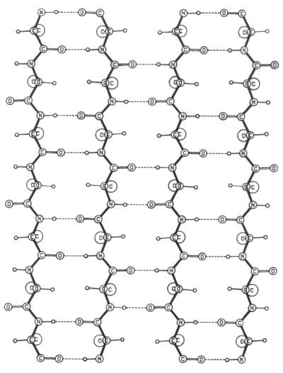
Pauling and Corey's alpha and beta models
Astbury was of course interested in the globular proteins that are
responsible for the body's various metabolic functions and he envisaged that
the protein chains in them would be folded back and forth in a regular
fashion, for example in the form of uniform stacks of sheets or ribbons.
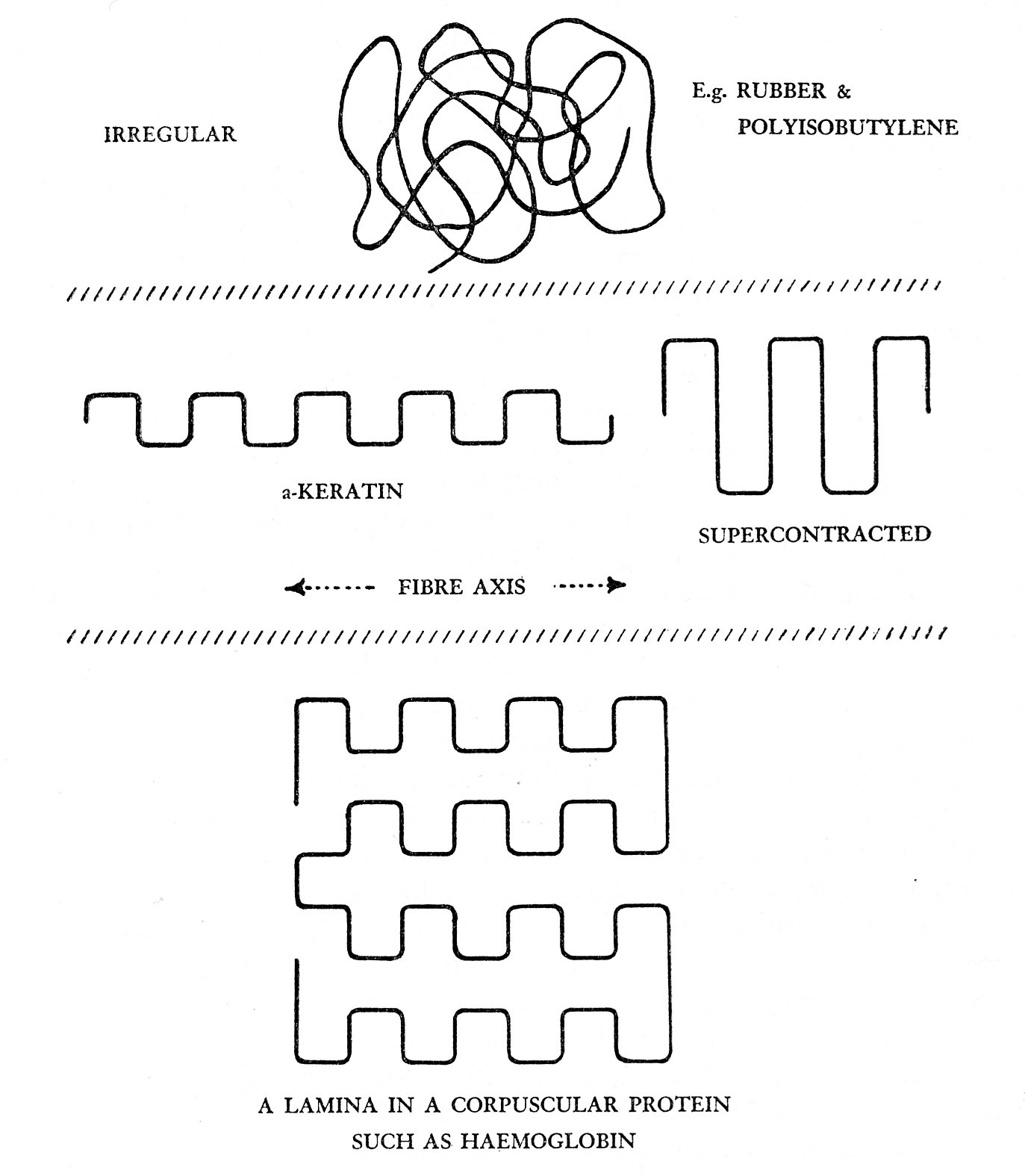
Here again, while the essence was right, the detail was not.
W L Bragg in Cambridge and London
In 1937, WLB moved from Manchester to be Director of the National Physical
Laboratory, but his stay there was short-lived, because Sir Ernest
Rutherford died unexpectedly and WLB was invited to succeed him in 1938 as
Cavendish Professor in Cambridge.
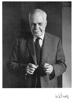
Hardly had he arrived when war delayed his full-time attention to his
department, but he had already been excited by the discovery by J D Bernal
and Dorothy Crowfoot (later Hodgkin) that globular proteins could be
crystallised provided they were prevented from drying out. Another arrival
in Cambridge had been Max Perutz, enthusiastic to find the 3-dimensional
structure of haemoglobin. WLB managed to obtain funding from the Medical
Research Council to support work on globular proteins, eventually giving
rise to the MRC Laboratory of Molecular Biology, probably the most
successful laboratory in the world, certainly as measured by Nobel Prizes
won by its members. Perutz was joined by John Kendrew, and together they
were responsible for the 3-dimensional structures of the first two globular
proteins, haemoglobin (the oxygen-carrier) which has 4 similar chains and
myoglobin (the oxygen-storage protein in muscle), a simpler molecule with
just one chain, like those in haemoglobin.
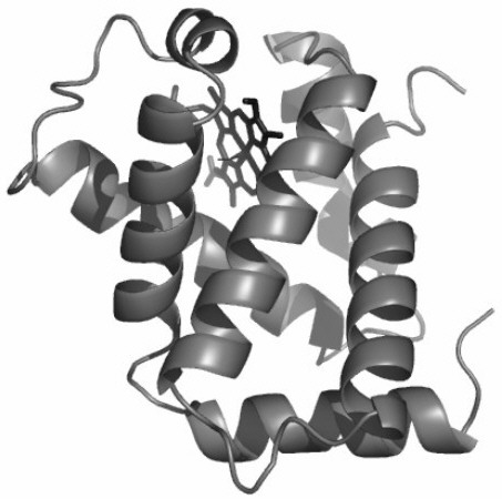
In each of the myoglobin and haemoglobin chains, an oxygen molecule
is carried by the iron atom at the centre of the flat haem group, which is
enfolded by the protein chain (right).
The myoglobin chain and each of the two different types of haemoglobin
chains are composed of about 8 segments, each in the form of alpha helices.
As Astbury had suggested, this globular protein consists of one of the
typical fibrous protein conformations folded into a compact near-spherical
shape - but the overall fold was nothing like as regular as he had
envisaged.
|
|
As the work on haemoglobin and myoglobin was progressing, WLB made the final
move of his career. Just as it had done before his father had moved there,
the Royal Institution was again in need of a firm directing hand, and WLB
was persuaded to take over there in 1955. (WHB had died over 10 years
previously.) However, WLB did not sever his links with Cambridge and he
obtained support from the Medical Research Council to appoint new research
staff at the R.I. who initially worked closely with Perutz and Kendrew on
determining the haemoglobin and myoglobin structures.
With those projects having reached fruition, the R.I. team went on to obtain
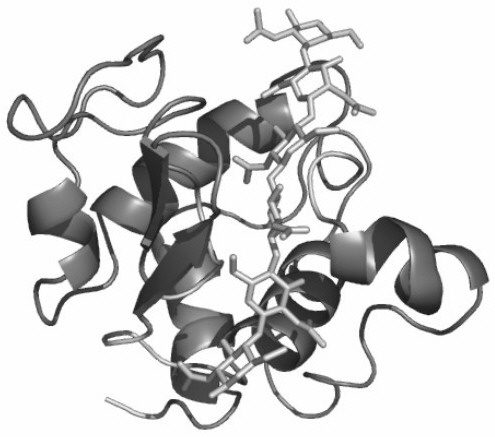
the structure of the third globular protein, lysozyme, an anti-bacterial
enzyme whose function is to break down bacterial cell walls by cutting a
polysaccharide component of the bacterial cell wall; the enzyme can bind 6
rings of the polysaccharide and cuts the chain between the 4th and 5th ring
from the top..
This was the first enzyme of known 3-dimensional structure, which permitted
the catalytic mechanism to be deduced. Its structural interest, however, is
that the lysozyme molecule is again an irregular shape, but, while some
parts of the chain have a helical fold, others are in the form of elongated
beta-type strands.
|
|
|
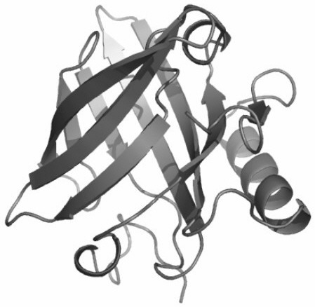
The occurrence of alpha and beta folds in different proportions in globular
proteins is a general property; in some cases the beta conformation
predominates, as in the mouse major urinary protein, (below right) a
component of the urine of male mice, which carries a pheromone
molecule that acts on female mice. It is a member of the lipocalin
family, several of which structures have been determined in Leeds in recent
years.
These two structural motifs are to be found in the very large protein
molecules and in macromolecular complexes such as viruses - work on a number
of such systems is in progress in Leeds, led nowadays by Simon Phillips and
colleagues in the Astbury Centre.
|
Astbury on protein denaturation
Globular proteins can quite easily be 'denaturated' by heat or chemical
attack into a random, unfolded form. Astbury was fascinated by the fact that
fibres could be formed from denatured globular proteins, and on wearing a
pullover made of a yarn spun from a globular protein, he wrote (in 1936):
"Only the other evening, I was watching my daughter knitting yarn spun
from monkey-nut protein - a protein, I repeat, with round molecules that
once seemed to bear no relation whatsoever with fibres - and as I touched
the knitting, again the wonder of it all flooded over me".
This sort of phenomenon has recently taken on great medical importance as it
has now become apparent that a number of diseases such as Alzheimer's,
Creutzfeld-Jacob's disease and Type II diabetes are associated with the
misfolding of protein chains - most remarkably into the 'cross-beta' type of
fold that Astbury's group had first observed. Work on protein folding and
misfolding is now being pursued in Leeds by Sheena Radford and her group.
Astbury's ideas on nucleic acids
DNA can be extracted from cells into a gel form, from which it is possible
to draw out fibres in which the molecules are aligned. Members of Astbury's
group took X-ray pictures of such fibres which showed a regular axial repeat
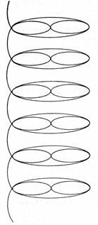
of 3.34Å. Astbury recognised this spacing as being similar to the
thickness of flat molecules such as graphite and benzene, and he knew that
the 4 types of DNA 'base' (A,T,G and C) are just such flat molecules; he
therefore deduced that DNA bases are stacked on top of each other "like a
pile of pennies".
He also thought that the similarity to the periodicities in fibrous protein
structures was significant, but in this respect he was wrong.
|
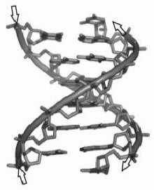
|
| Astbury's 'pile of pennies' model |
Crick & Watson's model |
Unfortunately for Astbury, for a variety of reasons, he did not take his DNA
work any further and it was in WLB's lab. in Cambridge where the essential
aspects of DNA structure were discovered by Francis Crick (who was supposed
to be working on haemoglobin) and James Watson (a young American visitor).
They deduced from the X-ray data that DNA molecules comprised two
anti-parallel chains, like the stiles of a ladder, linked together by pairs
of bases, A with T and G with C, which formed the rungs. Their
double-helical model showed directly how this complementary base-pairing
provided the basis of replication. The molecules can be copied exactly- if
the 2 strands are pulled apart; new strands can only be assembled if the
bases on each new "daughter" strand are again complementary to those of each
"parent".
Crick and Watson's model depended entirely on X-ray data obtained by Maurice
Wilkins and his colleagues (including Rosalind Franklin) in J T Randall's
department at King's College, London, and the systematic 'refinement' of the
model by Wilkins's group proved the correctness of the model. A further
interesting connection is that Randall had been a student of WLB in
Manchester.
Origins of the term 'Molecular Biology'
It seems that the first published use of the term 'Molecular Biology' was by
Warren Weaver, director of the Rockefeller Foundation's natural sciences
programme, in 1938: "Among the studies to which the [Rockefeller] Foundation
is giving support is a series in a relatively new field, which may be called
molecular biology, in which delicate modern techniques are being used to
investigate ever more minute details of certain life processes."
Astbury gave his own definition in 1961: "Molecular biology implies not so
much a technique as an approach, an approach from the viewpoint of the
so-called basic sciences with the leading idea of searching below the
large-scale manifestations of classical biology for the corresponding
molecular plan. It is concerned particularly with the forms of
biological molecules and .... is predominantly three-dimensional and
structural - which does not mean, however, that it is merely a
refinement of morphology - it must at the same time enquire into genesis and
function."
Nowadays, the term has a rather wider connotation in that much molecular
biology no longer has an explicit 3-dimensional structural quality, although
a 3-dimensional structural under-pinning is implicit.
Astbury himself, when a chair was conferred on him, would have liked the
title 'Professor of Molecular Biology'; unfortunately a senior Leeds
biologist expressed the view that, while Astbury knew a lot about molecules,
he did not know any biology, so Astbury was given the title 'Professor of
Biomolecular Structure'. I think that the criticism was unfair as, while
without a training in biology, Astbury was always aware of the biological
implications of his work.
Astbury's conception of his role in science
In the opinion of his biographer, J D Bernal, Astbury was responsible for
Leeds becoming the major centre of fibre research worldwide for more than 15
years following his appointment. He established the relationships between
the gross (anatomical) structure of natural materials, their physical
properties and their underlying molecular structures.
Astbury himself must have realised that, although he had laid the
foundations in his derivation of relationships between structure at the
molecular level and physical properties at the anatomical level, he had not
succeeded in deriving the structural details of the major classes of
biological macromolecules; he used the following quotation to illustrate
what he thought his role had been:
"The which observ'd, a man may prophesy,
With a near aim, of the main chance of things
As yet not come to life, which in their seeds
And weak beginnings lie intreasured."
Shakespeare, King Henry IV, part 2)
Summary
I should like to end with a further quotation:
"Did we know the mechanical affections of the particles of
rhubarb, hemlock, opium and a man, as a watchmaker does
those of a watch...............we should be able to tell
beforehand that rhubarb will purge, hemlock kill and opium
make a man sleep................"
(John Locke in his Essay Concerning Human Understanding (1620))
I would claim that the work of the Braggs in 1912-1913 laid the foundations
of X-ray crystallography as a method for the determination of the structures
of materials and especially of those of biological origin. Astbury's work,
from his arrival in Leeds in 1928 and using X-ray methods, was seminal in
developing our understanding that the properties of living organisms could
be understood from a knowledge of the structures and properties of their
constituent molecules.
Through the work of these men and those who have followed them, we have
progressed a long way to achieving Locke's aspiration.
Sources of material
- Biographical Memoirs of Fellows of the Royal Society:
- J D Bernal: William Thomas Astbury
- Sir David Phillips: William Lawrence Bragg
- G M Caroe: William Henry Bragg
- John Jenkin: The Bragg Family in Adelaide
- Robert Olby: The Path to the Double Helix
- Graeme Hunter: Light is a Messenger
(The life and science of William Lawrence Bragg)
Next
Top


